Home

živ taksi Montgomery canalis spiralis Marquee kanal Ždrijelo

Clinical anatomy of the upper extremity

Fascija | Facebook
Fascija | Facebook

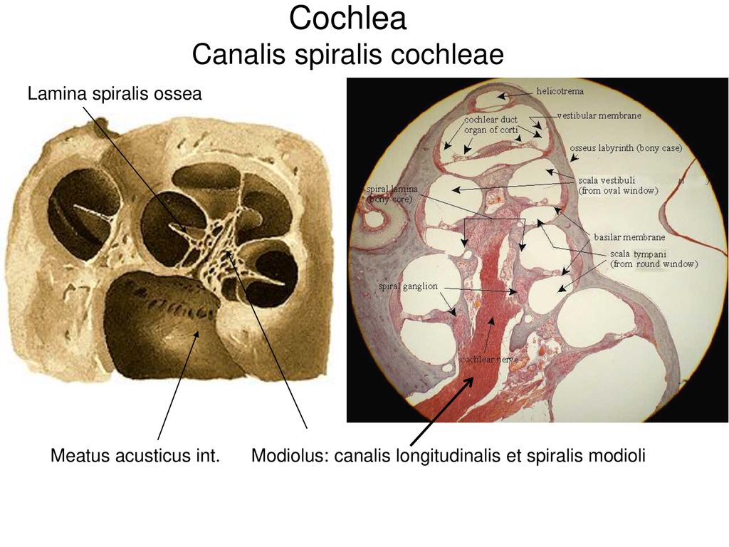
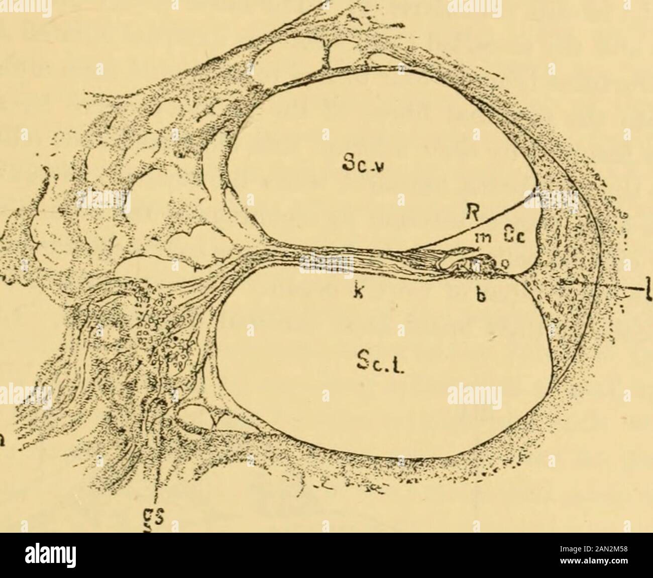
A text-book of the diseases of the ear and adjacent organs . amina spiralis  ossea obliquely to the external wall of thecochlea, into two divisions, of  which the one formed by the
A text-book of the diseases of the ear and adjacent organs . amina spiralis ossea obliquely to the external wall of thecochlea, into two divisions, of which the one formed by the

Topografia do membro superior Diagram | Quizlet

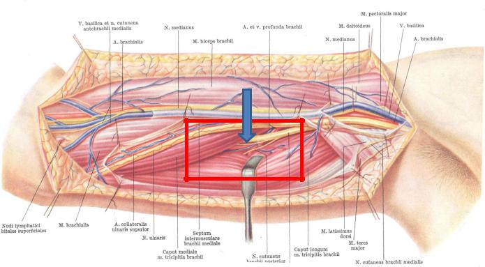
Canalis humeromuscularis (seu canalis spiralis seu canalis n.radialis) The  canal walls: 1. Sulcus.. | VK
Canalis humeromuscularis (seu canalis spiralis seu canalis n.radialis) The canal walls: 1. Sulcus.. | VK
M.Rogha M.D Isfahan university of medical sciences - ppt video online  download
M.Rogha M.D Isfahan university of medical sciences - ppt video online download

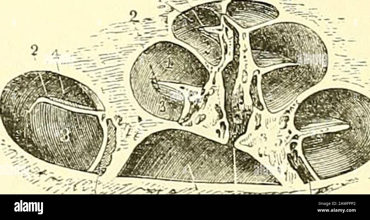
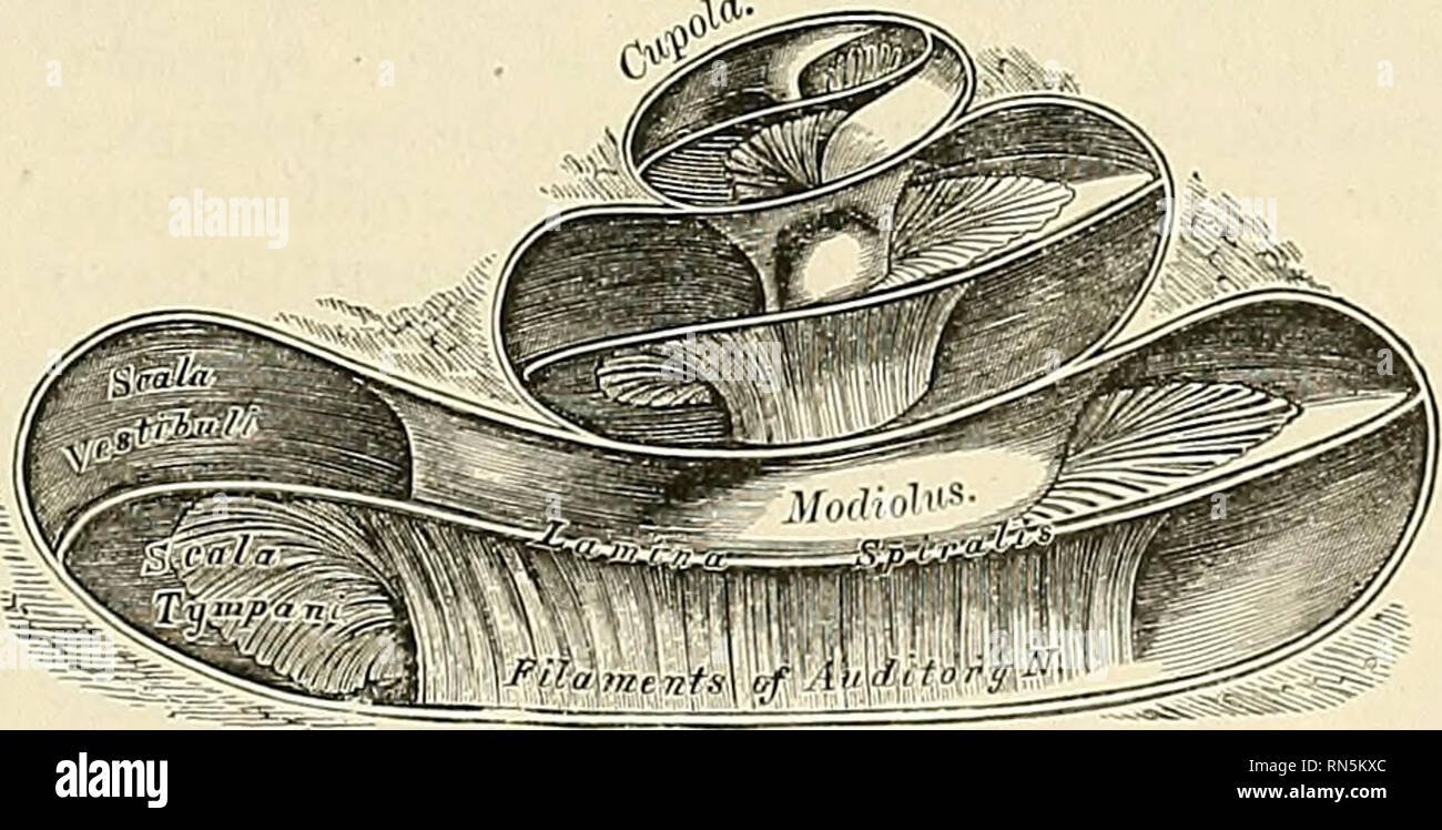
Quain's elements of anatomy . Fig. 388.—Diagrammatic view of the osseous  cochlea laid open, s1, modiolus or central pillar ; 2, placed on three  turns of the lamina spiralis ; 3, scalatympani ;
Quain's elements of anatomy . Fig. 388.—Diagrammatic view of the osseous cochlea laid open, s1, modiolus or central pillar ; 2, placed on three turns of the lamina spiralis ; 3, scalatympani ;

Section Through Coil of the Cochlea | ClipArt ETC

Canalis humeromuscularis (seu canalis spiralis seu canalis n.radialis) The  canal walls: 1. Sulcus.. | VK
Canalis humeromuscularis (seu canalis spiralis seu canalis n.radialis) The canal walls: 1. Sulcus.. | VK
Fascija | Facebook
Fascija | Facebook

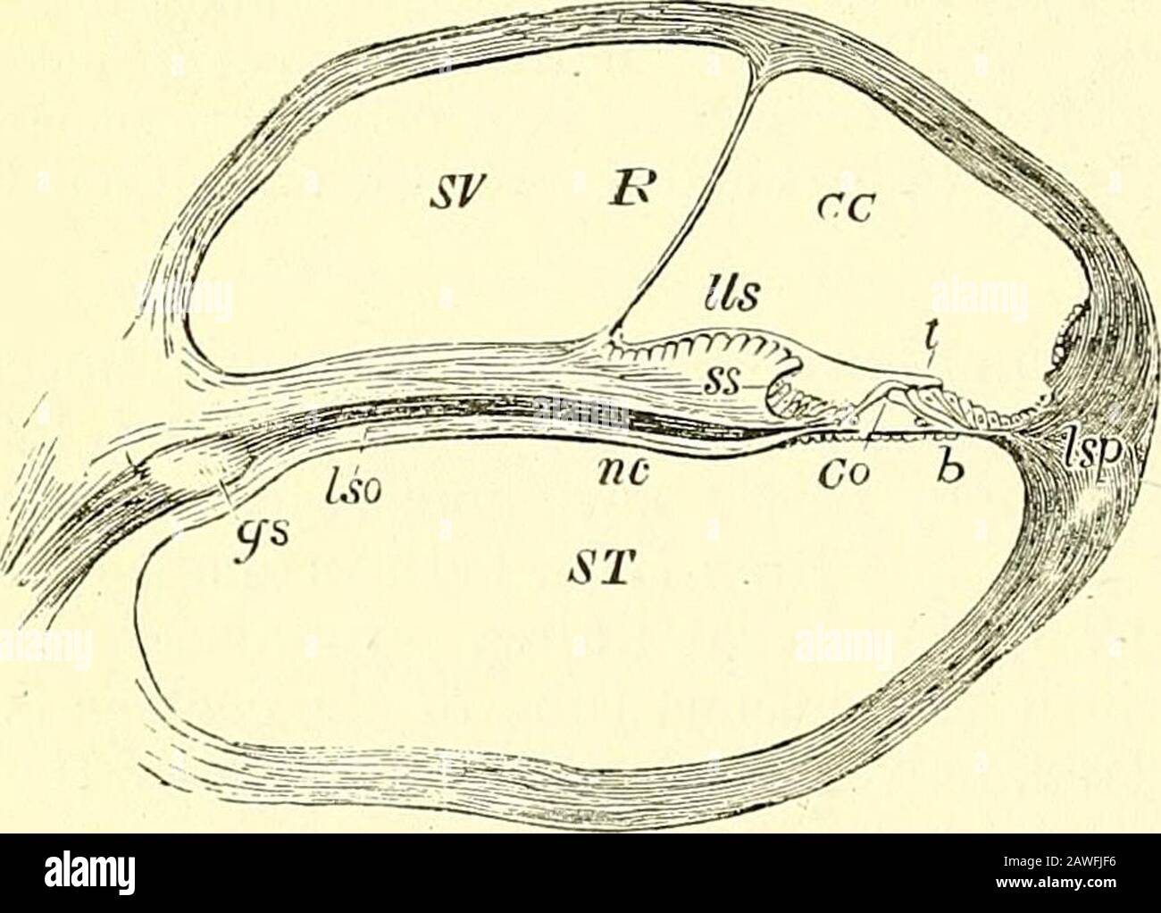
Quain's elements of anatomy . amina spiralis. scala tympani (ST), but does  not properly speaking enter into the lowerboundary of the scala vestibuh,  for a second, much more delicate mem- Fig. 398.
Quain's elements of anatomy . amina spiralis. scala tympani (ST), but does not properly speaking enter into the lowerboundary of the scala vestibuh, for a second, much more delicate mem- Fig. 398.

Organum vestibulocochleare Auris interna, hallórendszer - ppt letölteni

Clinical anatomy of the upper extremity

Caroticotympanic canals | definition of caroticotympanic canals by Medical  dictionary
Caroticotympanic canals | definition of caroticotympanic canals by Medical dictionary

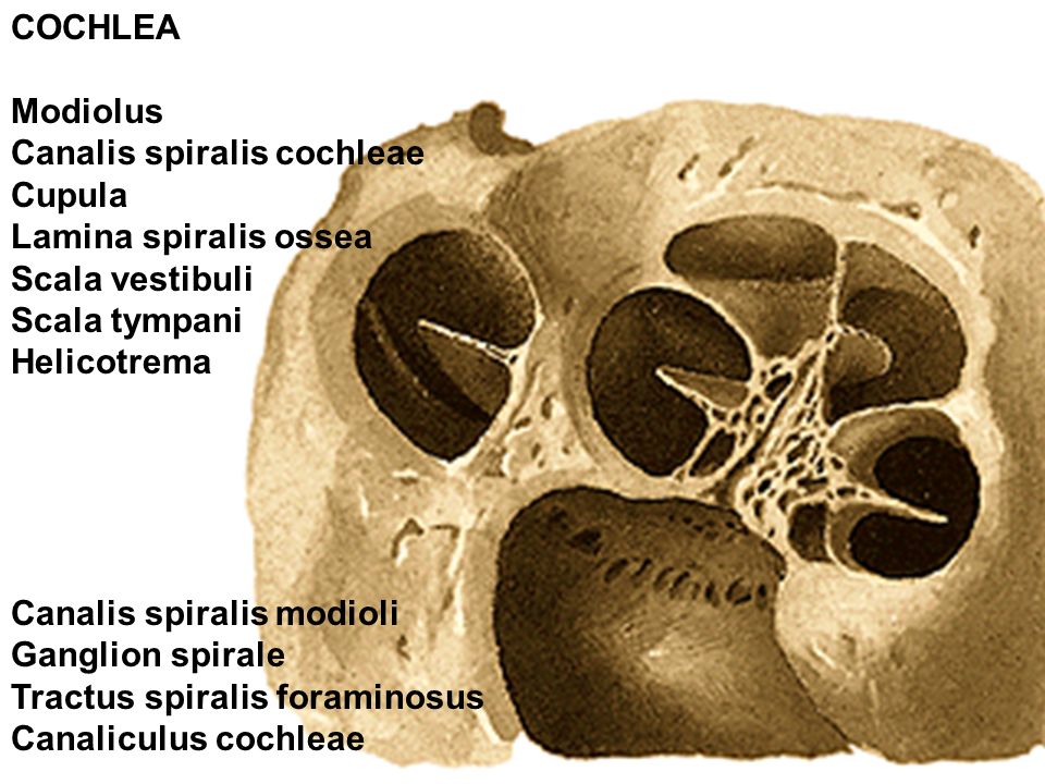
Anatomy, descriptive and applied. Anatomy. LAMINA SPIRALIS OSSEA Fig.  858.—Vertical section through the right cochleo, medial portio 1 the  lateral side. (Spalteholzj. foramen centrale. The foramina of the tractus  spiralis foraminosus
Anatomy, descriptive and applied. Anatomy. LAMINA SPIRALIS OSSEA Fig. 858.—Vertical section through the right cochleo, medial portio 1 the lateral side. (Spalteholzj. foramen centrale. The foramina of the tractus spiralis foraminosus
Introduction. History of otorhinolaryngology - ppt video online download

Презентация на тему: Отверстия задней стенки подмышечной полости

the human ear
the human ear

Image from page 988 of "The comparative anatomy of the dom… | Flickr

Anatomy, descriptive and applied. Anatomy. LAMINA SPIRALIS OSSEA Fig.  858.—Vertical section through the right cochleo, medial portio 1 the  lateral side. (Spalteholzj. foramen centrale. The foramina of the tractus  spiralis foraminosus
Anatomy, descriptive and applied. Anatomy. LAMINA SPIRALIS OSSEA Fig. 858.—Vertical section through the right cochleo, medial portio 1 the lateral side. (Spalteholzj. foramen centrale. The foramina of the tractus spiralis foraminosus

Canalis humeromuscularis (seu canalis spiralis seu canalis n.radialis) The  canal walls: 1. Sulcus.. | VK
Canalis humeromuscularis (seu canalis spiralis seu canalis n.radialis) The canal walls: 1. Sulcus.. | VK
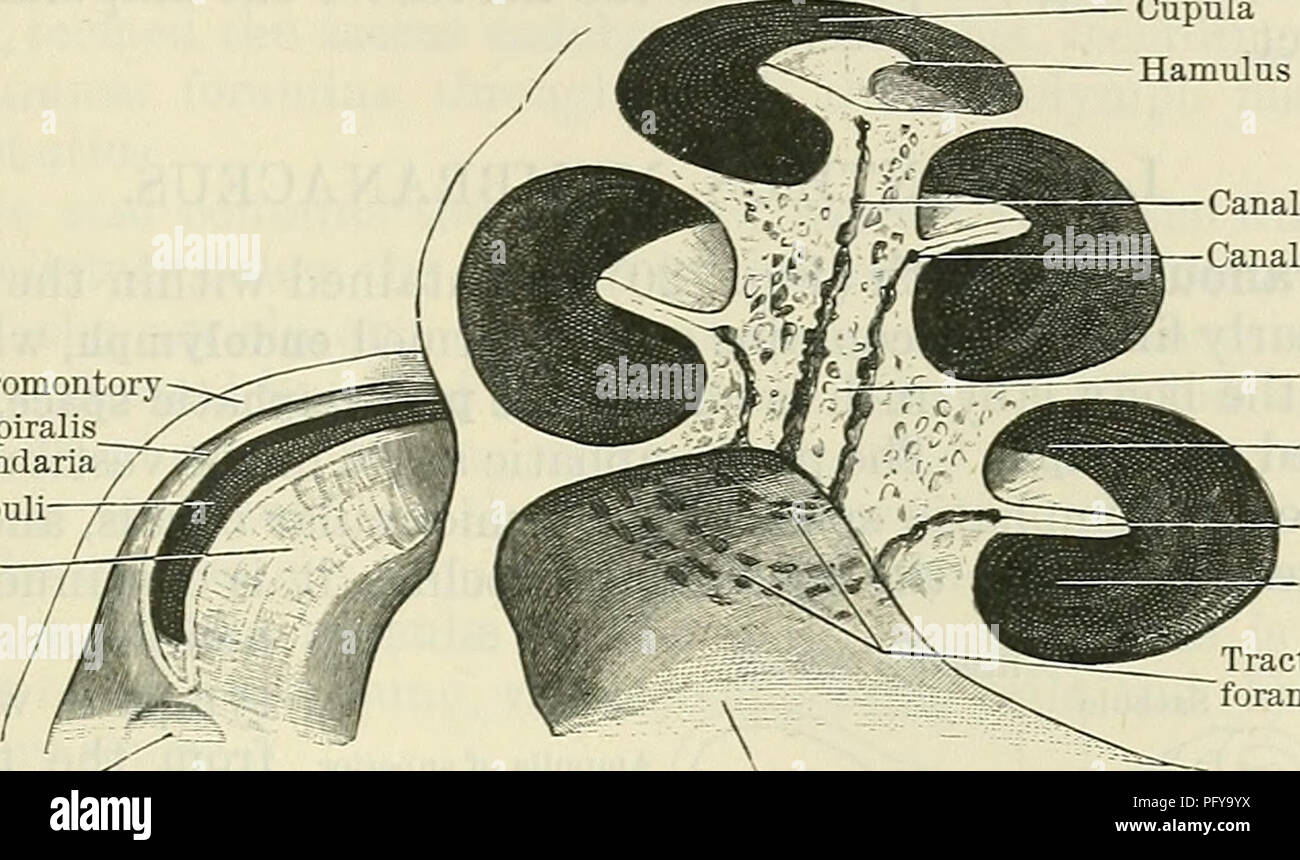
Cunningham's Text-book of anatomy. Anatomy. OSSEOUS LABYKINTH. 845 varying  distance, they bend outwards to reach the attached edge of the lamina  spiralis ossea, where they expand and form by their apposition
Cunningham's Text-book of anatomy. Anatomy. OSSEOUS LABYKINTH. 845 varying distance, they bend outwards to reach the attached edge of the lamina spiralis ossea, where they expand and form by their apposition

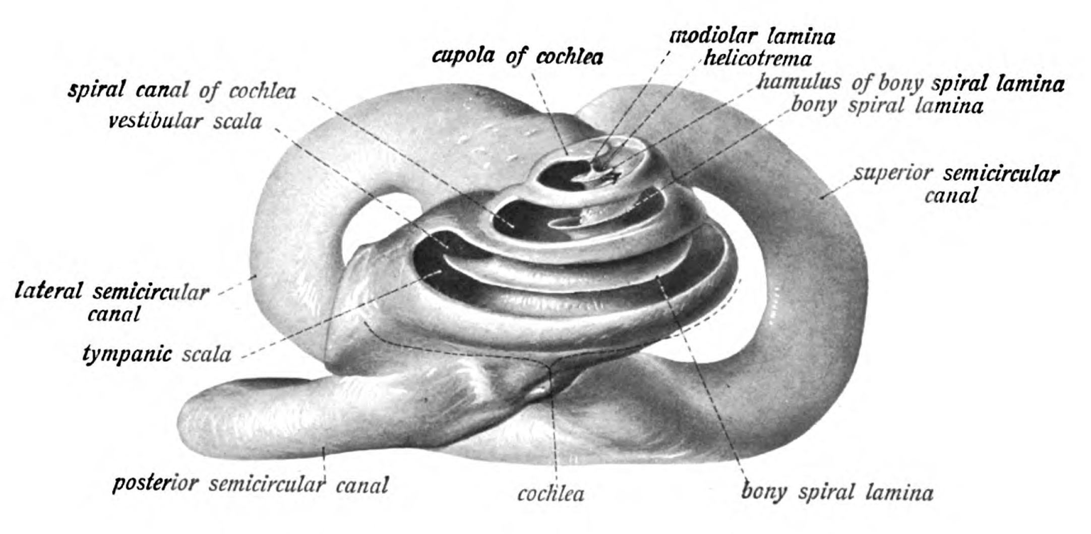
Sobotta 1911 fig.786 - Bony labyrinth, median/anterior view - English  labels | AnatomyTOOL
Sobotta 1911 fig.786 - Bony labyrinth, median/anterior view - English labels | AnatomyTOOL

Cochlear duct - Wikipedia

Section of cochlea vintage Royalty Free Vector Image

The Ear BG
The Ear BG

Osseous spiral lamina - Wikipedia

ORGANUM VESTIBULOCOCHLEARE. - ppt video online download